Malignant melanoma is a less common but highly dangerous skin cancer.

When found at an early stage, melanoma has one of the highest cure rates of all cancers, at more than 90 per cent. If left untreated, melanoma will invade the skin. When it reaches the bloodstream or the lymphatic system, it can spread to other parts of the body and often causes death.

What is it?

Melanoma is a form of skin cancer. It starts in the melanocytes or pigment-producing cells in the outer layer of the skin. These cells grow out of control and form a tumour. Melanomas are often brown and black, but can also appear in other colours.

Who gets it?

In 2019, melanoma was the 7th most common type of newly diagnosed cancer for both males and females. Among youth and young adults (ages 15 to 29), melanoma was the 4th most common newly diagnosed cancer – 7% of the total new cases diagnosed – after thyroid, testicular, and Hodgkin lymphoma.

Approximately 8,000 Canadians will be diagnosed with melanoma in 2020, and 1,300 will die of it. While the incidence rates of some types of cancer are decreasing every year (e.g. laryngeal for both sexes, lung cancer in males, and cervical cancer in females), melanoma is among the types that continue to increase annually.

IN MALES, the incidence rate for melanoma has increased steadily at 2.2% per year since 1984 – the 3rd-largest overall increase in incidences for males, after thyroid cancer (6.4%) and multiple myeloma (2.6%).

IN FEMALES, the incidence rate for melanoma was stable from the mid-1980s to the mid-1990s; since 1994, the rate has increased by 2.0% per year – the 2nd-largest overall increase in incidences for females, after liver cancer (2.7%).

Studies show older, fair-skinned men are more likely to die of melanoma than any other group. A lack of awareness of this disease and its frequent location in a hard-to-see area – the back – are likely the main reasons.

In Canada, the lifetime risk of melanoma for men is now 1 in 59. For women, it is 1 in 73. In comparison, the lifetime risk of melanoma for North Americans in the 1930s was 1 in 1,500.

What causes it?

Excessive exposure to ultraviolet (UV) from the sun and tanning beds plays a leading role in the development of melanoma and is the most preventable cause. Experts estimate about 90 per cent of melanomas are associated with severe UV exposure and sunburns over a lifetime.

How can I protect myself?

The best ways are:

  • Find out your risk — the risk factors are well known.
  • Learn the early signs of melanoma (see the Skin Cancer Self-Examination poster); find out why and how to check your skin.
  • Protect yourself from the sun from spring to fall, and avoid tanning beds.
  • Check your skin monthly.
  • Consult a doctor if you see any suspicious spots.
Early detection linked to survival 

Unlike many cancers, melanoma is clearly visible on the outer surface of the skin. In the early stages the tumour is thin, has not spread to lymph nodes and is curable by surgical removal.

Risk factors?

People more likely to develop melanoma have:

  • fair, sun-sensitive skin that burns rather than tans; freckles; red or blond hair
  • many moles — more than 50
  • moles that are large or unusual in colour or shape
  • close family history of melanoma, or a personal history of melanoma
  • excessive exposure to UV from the sun or  tanning bed
  • history of severe sunburns

The risk is multiplied if you have several of these risk factors, for example, if you have unusual moles and a family history of melanoma.

People with no risk factors, and those with darker skin, can also get melanoma.

Why should I check my skin?
  • People are very successful at detecting melanoma on their own skin or that of a family member. Research shows that 53 per cent of melanomas are discovered by the patients themselves, and a further 17 per cent by family members.
  • Checking your skin can lead to early detection and improved survival. Early stage melanomas have more than 90 per cent cure rate. Lives can be saved.
  • A skin self-exam is simple, requiring just 10-15 minutes once per month.
  • Recent research shows those at risk who had a friend or family member help with checking the skin found the disease at a much earlier stage, and had a 63 per cent lower death rate, compared with those who did not check their skin.
  • Having a history of melanoma increases the risk of developing melanoma and non-melanoma skin cancer.
  • There is a  risk of melanoma recurrence if disease found in advanced stages
What does melanoma look like?

Melanoma can develop in weeks, months or years. It can appear as a new mole or freckle-like spot on the skin, or develop in an existing mole. Melanomas are usually dark brown or black, although some show a mixture of colours including blue, grey and red.

The typical location for melanoma in men is on the back; in women, it’s the leg. It can also appear on the arm, scalp or face. While less common in darker-skinned people, melanoma can appear on soles of the feet, toenails and palms of the hands.

The ABCDE of melanoma will help you to detect this disease. Look for these features:

ABCDE of Melanoma

Asymmetry – The shape on one side is different from that on the other side

Border – The border or visible edge is irregular, ragged and imprecise

Colour – There is a colour variation, with brown, black, red, grey or white within the lesion

Diameter – Growth is typical of melanoma. It can measure more than 6 mm, although it can be less

Evolution – Look for change in colour, size, shape or symptom, such as itching, tenderness or bleeding

The ugly duckling sign

This tip may help you detect melanoma. Most moles look similar. However, melanomas look different from all other moles — the ugly duckling sign. Generally only one melanoma appears at a time, so a spot that looks or feels different, or changes differently compared to other moles on the body, should be checked by your dermatologist or family doctor as soon as possible.

What if a suspicious spot or mole is found?

See your dermatologist or family doctor as soon as possible to have the lesion examined. A skin biopsy under local anesthesia may be needed. A pathologist will then examine the sample.

What if melanoma is found?

If the biopsy confirms melanoma, further surgery will be needed to remove a safety margin around the site. This serves to remove cancer cells that may still exist at the melanoma site.

A cancer diagnosis can sometimes be a significant emotional shock. Patients are advised to express their concerns about the melanoma diagnosis; some may occasionally need help from professional counsellors.

Factors affecting treatment

The main factor in determining treatment is the depth of the melanoma in the skin, as measured by the pathologist (Breslow index). The deeper the cancer, the more likely it has spread and the more likely it will recur.

Melanomas less than 1 mm thick rarely spread. Removal of the melanoma and a surrounding margin of skin completes treatment. Other factors affecting treatment include location of the melanoma and ulceration.

Further tests

If the melanoma is thicker (usually more than 1 mm), a lymph-node biopsy is often suggested to test for cancer cells in the lymph node closest to the melanoma. The results help doctors determine the stage of melanoma and the treatment needed. Such a biopsy is often taken during surgery to remove the melanoma. Blood tests, a CT scan or a nuclear-medicine examination called a PET-scan may be used. (See Stages of melanoma.)

Types of Melanoma

There are four main types:

Superficial spreading melanoma accounts for about 70 per cent of cases. These lesions tend to grow outwards, so watch for spots or moles that are growing/spreading.

Nodular melanoma is a more dangerous form of melanoma since it grows quickly downward into the skin. It often appears on areas not usually exposed to the sun. It appears as a raised area and is usually very dark, but sometimes reddish.

Lentigo maligna melanoma appears on the parts of the body constantly exposed to direct sunlight. It is frequently found among older people. It often appears as a fairly wide, flat brown patch marked by several hues of brown and black.

Acral lentiginous melanoma is the most common melanoma in dark-skinned people and those of Asian descent. It appears on soles of feet, palms of hands and under toenails and fingernails.

Stages of Melanoma

There are five stages of melanoma based on the thickness and other features of the tumour. These stages provide an important guide to treatment, indicate the risk of recurrence and determine whether other tests are needed.

Stage 0

Melanoma in situ is the most frequent stage, when the tumour is limited to the outer layer of the skin and has not spread. Surgery is done to remove the melanoma and the surrounding skin, which completes the treatment. The prognosis is excellent.

Stage 1

These are very early melanomas. Depending on the severity, a sentinel lymph-node biopsy is sometimes suggested. Treatment also includes a local re-excision.

Stage 2

The tumour is more advanced and is deeper in the skin. A sentinel lymph-node biopsy is usually suggested. After surgery there is a moderate risk of recurrence or spread to another part of the body because of the depth of the tumour.

Stage 3

The melanoma has spread to nearby lymph nodes; these are removed in treatment.

Stage 4

This is an advanced stage of melanoma. The cancer has spread to another part of the body such as the lungs, liver, brain or abdomen. This situation is rare.

For Stages 3 and 4, post-surgical treatment focuses on preventing the cancer from coming back and on killing the cancer cells in the body. The following treatments, used alone or in combination, may be prescribed.

Chemotherapy 
Chemotherapy drugs are often used to treat local recurrence, advanced melanoma that has spread or to control the symptoms of advanced melanoma. Side-effects can include nausea and vomiting, fatigue and hair loss. Chemotherapy can also increase the risk of infection for a time after treatment.

Biological therapy (or immunotherapy)
Biological therapy boosts the immune system to help the body act against cancer cells. These drugs target specific types of cells and, less frequently, damaged normal cells. Side-effects of biological therapy can include fatique, fever or chills, rashes or reactions at the injection site. Biological therapies used to treat melanoma include interferon and interleukin-2, ipilimumab, nivolumab and pembrolizumab

Targeted therapy
Targeted therapy involves treatment for patients whose melanoma has specific genetic changes such as a BRAFV600 mutation. Such mutations are found by testing samples of a patient’s tumour. The MEK gene works together with BRAF gene. MEK inhibitors help BRAF inhibitors to fight tumor cells. Currently approved targeted therapies in Canada include vemurafenib, dabrafenib and trametinib. Some side-effects of targeted therapy may include skin toxicities, fevers and chills, eye problems, diarrhea and heart problems. Several new targeted therapies are being evaluated in clinical trials.

Radiation therapy
Radiation therapy uses high energy x-rays or other types of radiation to kill cancer cells or stop them from growing. It is sometimes used after surgery to relieve or control symptoms, or to treat melanoma that has spread to the brain.

Radiation therapy damages both normal cells and cancer cells in its path. Side-effects depend on the area treated, and may include skin redness or irritation and fatigue. In stage III melanoma, radiation may be used after surgery to prevent spread and recurrence. In stage IV disease, radiation may help control symptoms.

5 Step Skin Cancer Self-Examination

While people may be unsure how best to check their skin, performing a self-examination using the steps below and the “ABCDEs” is a simple way to remember the key characteristics that could identify a potential case of melanoma.

Self-exam step 1 image 
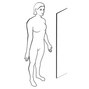
Using a mirror in a well-lit room, check the front of your body – face, neck, shoulders, arms, chest, abdomen, thighs and lower legs.
Self-exam step 2 image 
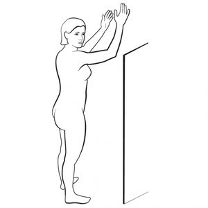
Turn sideways, raise your arms and look carefully at the right and left sides of your body, including the underarm area.
Self-exam step 3 image 
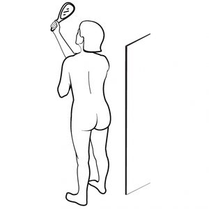
With a hand-held mirror, check your upper back, neck and scalp. Next, examine your lower back, buttocks, backs of thighs and calves.
Self-exam step 4 image 
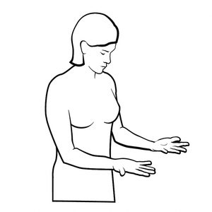
Examine your forearms, palms, back of the hands, fingernails and in between each finger.
Self-exam step 5 image 
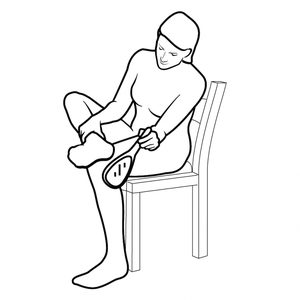
Finally, check your feet – the tops, soles, toenails, toes and spaces in between.